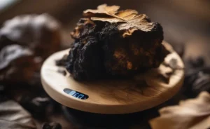

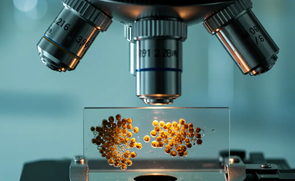
Much like appreciating a woven tapestry from a distance gives no hint of the intricate threads forming its pattern, the familiar exterior of a morel mushroom conceals a world of microscopic detail. For mycologists and enthusiasts, understanding this hidden dimension is essential for definitive identification and appreciating the fungal life cycle. Placing a sample of a morel mushroom under microscope reveals a fascinating landscape of spores, asci, and other cellular structures that define this prized edible. This detailed view provides invaluable information about the mushroom’s biology, from its reproductive strategies to the subtle characteristics that distinguish it from potentially toxic look-alikes. This guide explores the necessary equipment, preparation techniques, and the remarkable features you can expect to discover.
Morel Mushroom Under Microscope
Observing a morel mushroom at the cellular level provides definitive characteristics that visual inspection alone cannot offer. The unique pitted and ridged surface of the cap is a complex reproductive structure, and its true nature is only revealed through microscopy. This examination allows for the detailed study of the spores (ascospores) and the sacs that produce them (asci), which are fundamental for confirming the mushroom’s identity and distinguishing it from non-edible or toxic species. Studying morel mushroom identification requires this detailed approach.
Essential Microscope Equipment for Spore Viewing
To properly study morel spores and other microscopic features, a specific set of tools is required. The most crucial piece of equipment is a compound light microscope capable of various magnification levels. Other essential items include:
- Glass Slides and Coverslips: These are used to mount the specimen for viewing. A clean slide and a thin coverslip are necessary to create a clear field of vision.
- Mounting Medium: A drop of water is often sufficient for viewing spores. However, special stains can be used to highlight different cellular structures, making them easier to see.
- Sample Collection Tools: A sharp scalpel or a clean razor blade is needed to obtain a thin section of the mushroom tissue or to scrape spores from a spore print.
- Immersion Oil: For the highest levels of magnification, immersion oil is used between the objective lens and the coverslip to improve the resolution and clarity of the image.
Preparing Morel Spores for Microscopy
The first step in preparing a sample is often to create a spore print. This is done by placing the morel cap, pit-side down, on a piece of paper or glass for several hours. The cap will release its spores, leaving a visible print. A tiny amount of these spores can then be scraped onto a clean microscope slide. Add a single drop of water or mounting fluid over the spores, and then gently lower a coverslip over the drop, being careful to avoid air bubbles. An alternative method involves taking a very thin cross-section of the mushroom cap’s hymenium (the spore-bearing surface) and mounting it directly on the slide.
Magnification Levels for Morel Spores
Different levels of magnification are used to observe various features. A lower power setting, such as 100x, is useful for locating the spores on the slide and observing larger structures like the asci. To examine the details of individual spores, a higher magnification of at least 400x is necessary. The most detailed analysis, which allows for the observation of subtle surface textures and internal features of the ascospores, is best achieved at 1000x magnification with the use of immersion oil. Examining a morel mushroom under microscope at these varying levels provides a complete picture of its reproductive anatomy.
Microscopic Features of Morel Mushrooms
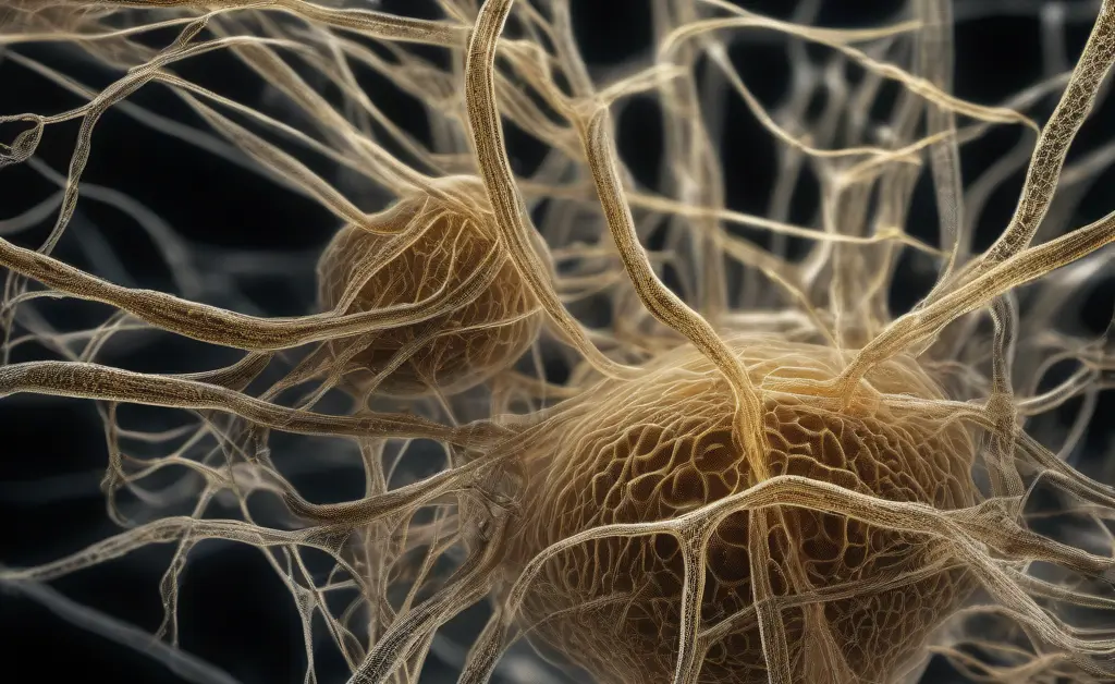
Beyond spores, the tissue of the morel itself presents a fascinating subject for microscopic study. The arrangement of its cells, known as hyphae, forms the mushroom’s structure and texture. When viewing a thin section of a morel mushroom under microscope, one can observe how these filamentous cells are interwoven to create the cap and stem. The fertile surfaces within the pits of the cap are distinct from the sterile ridges, a key characteristic of the *Morchella* genus. This detailed examination helps differentiate true morels from other fungi, as the cellular organization is unique. Understanding these morel mushroom characteristics is crucial for identification.
Microscopic Structures of the Morel Cap and Stem
The cap of a true morel is not a solid structure but a hollow network of pits and ridges. Microscopically, the surfaces inside the pits are the fertile areas, known as the hymenium. This layer is densely packed with elongated, sac-like cells called asci. Each ascus typically contains a set number of ascospores, which are the reproductive units of the fungus. Interspersed among the asci are sterile cells called paraphyses. In contrast, the ridges that form the network around the pits are sterile, composed entirely of interwoven hyphae and lacking any asci. The stem, or stipe, is also composed of hyphae, but their arrangement creates a different, more brittle texture than the cap. The study of morel mushrooms (Morchella spp.) reveals these intricate structures.
Observing Morel Mushroom Spores Under a Microscope
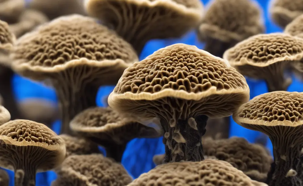
The spores are arguably the most important feature for definitive mushroom identification at the microscopic level. Morel spores are ascospores, meaning they are produced internally within an ascus. Observing their specific characteristics—shape, size, color, and surface texture—is critical. These features provide a reliable “fingerprint” that helps confirm whether a specimen is a true morel and can be used to distinguish it from its potentially dangerous look-alikes, such as those in the *Gyromitra* family. This distinction is vital for morel mushroom vs. false morel identification.
Spore Shape and Size
Morel ascospores have a distinct and consistent shape. They are typically smooth and broadly elliptical to oval. This uniform, simple shape is a key identifying feature of the *Morchella* genus. While their exact size can vary slightly between species, their general form remains constant. Observing this smooth, elliptical shape is a primary step in distinguishing them from the spores of other fungi, which may be spherical, angular, or ornately textured. This consistency is invaluable for mycologists.
Spore Coloration
When viewed as a thick deposit in a spore print, morel spores typically appear as a pale cream to yellowish or light buff color. However, when viewed individually under a microscope, the spores are hyaline, which means they are transparent and colorless. This is an important distinction, as the spore print color can be influenced by the density of the deposit. Under magnification, their lack of pigment is a consistent trait that helps in the identification process and contrasts with many other types of fungi whose individual spores may be darkly pigmented.
Key Differences in Spore Morphology
One of the most critical applications of examining a morel mushroom under microscope is differentiating it from toxic “false morels.” For example, species in the genus *Gyromitra* have spores that are markedly different. While true morel spores are smooth and elliptical, *Gyromitra* spores can be more rounded or fusiform (spindle-shaped) and often feature distinct ornamentation or one or more oil droplets inside. These differences in spore morphology are clear and definitive under high magnification. Verifying the presence of smooth, elliptical ascospores is a final, crucial step in confirming a mushroom’s identity as a true morel.
In conclusion, the practice of observing a morel mushroom under microscope transforms the identification process from a skill based on general appearance to a science based on concrete evidence. The cellular landscape it reveals—from the arrangement of hyphae in the cap and stem to the precise shape and texture of the ascospores—provides a level of certainty that macroscopic features cannot match. This microscopic view not only confirms the identity of the prized edible but also deepens our appreciation for the complex life cycle of fungi, reinforcing the importance of careful, detailed analysis in the field of mycology.
The content provided in this article is intended for informational learning, not clinical application. The information is not a substitute for professional mycological expertise. Never consume a wild mushroom unless you are absolutely certain of its identification, as many toxic species can resemble edible ones. Misidentification can lead to severe illness or be fatal. Always consult with a local expert before consuming any foraged fungi. Further research on fungal biology can be found at the NIH.
FAQ
What do morel mushroom spores look like?
Morel mushroom spores are typically elliptical or spindle-shaped and hyaline, meaning they are transparent. Under magnification, their surface can sometimes appear to have a fine reticulation or network pattern, though this varies by species. These microscopic features are important for identification.
What is the magnification for viewing spores?
Viewing mushroom spores, including those of the morel mushroom under microscope, generally requires a compound microscope. Magnifications ranging from 400x to 1000x are commonly used to clearly observe their size, shape, and surface characteristics.